√ club foot horse x ray 145335-Club foot horse x ray
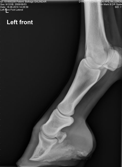
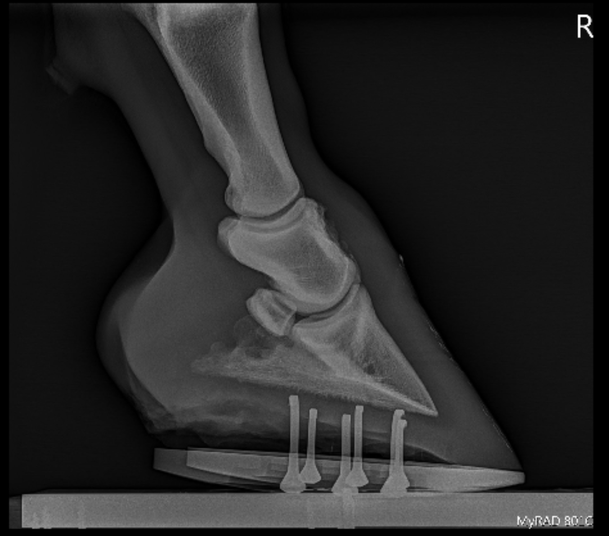
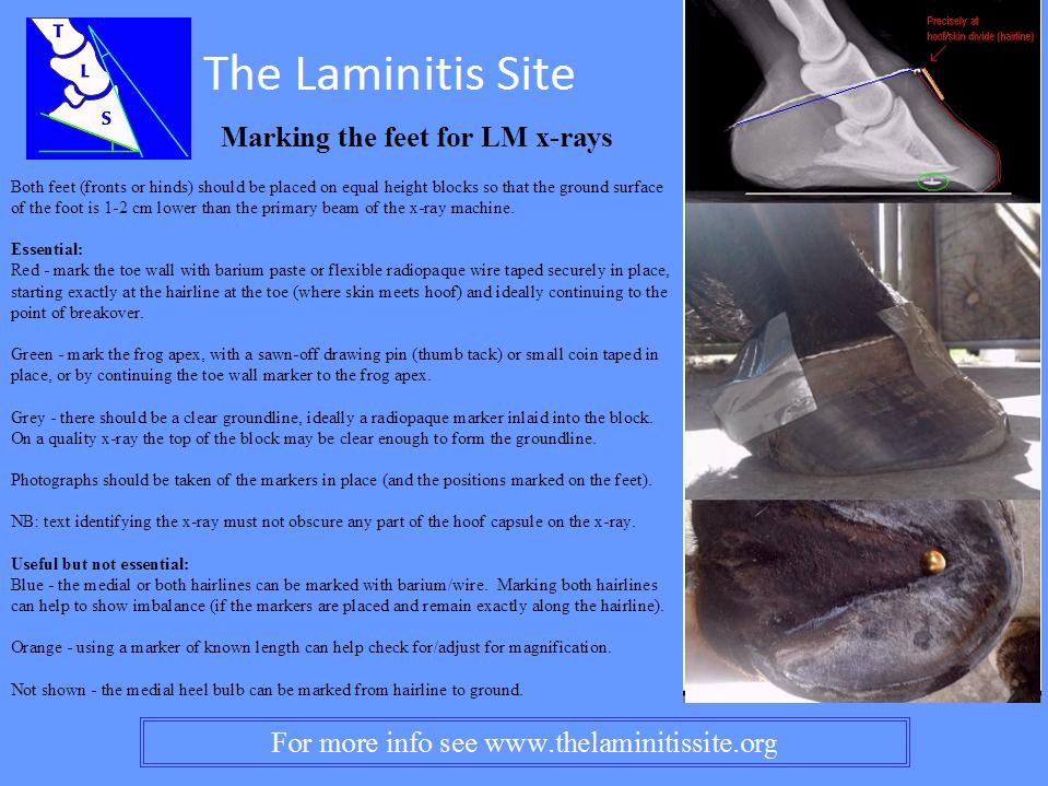
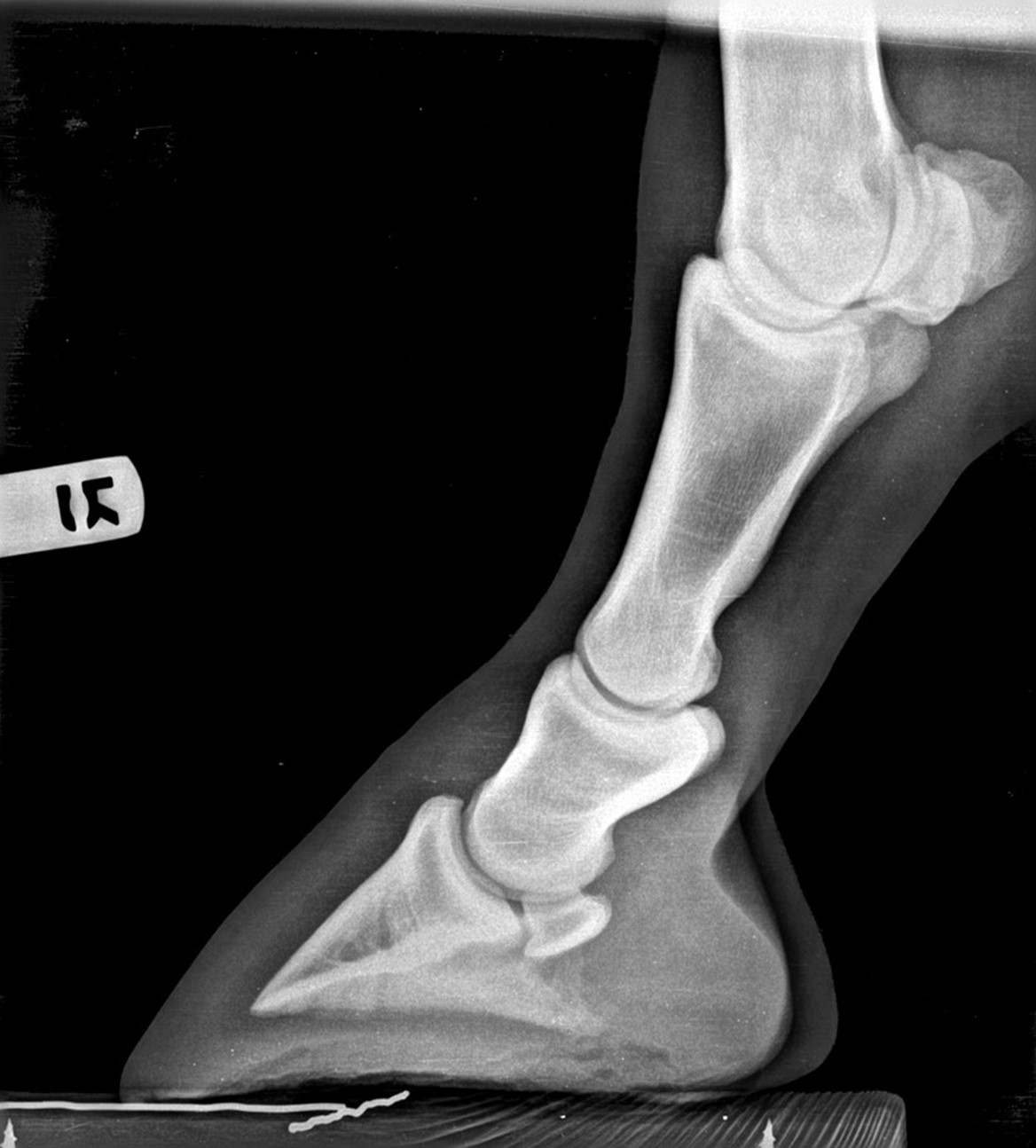
By Christy West, TheHorsecom Webmaster Article # 9805 When you look at a radiograph (X ray) of a horse's foot, do you visualize soft tissues, or do you only see bones?If possible, always take photographs of the foot in position at the time of the xray, showing the markers in place for correct interpretation of the xrays questions will be asked such as was the medial quarter of the hoof touching the cassette (if not, hoof wall thickness cannot be measured), was the horse equally weight bearing and were the cannon bones vertical (if not, bony columnVolar view with shoe on
Equine Podiatry Dr Stephen O Grady Veterinarians Farriers Books Articles
Club foot horse x ray
Club foot horse x ray-Equine Club Foot The equine club foot is defined as a hoof angle greater than 60 degrees What we see externally as the equine clubbed foot is actually caused by a flexural deformity of the distal interphalangeal joint (coffin joint) Causes include nutritional issues, heredity, position in the uterus orEquine radiography – the palmaroproximalpalmarodistal ('skyline') view of the distal sesamoidean ('navicular') bone A case study explaining the indications, preparations and technique required to carry out a radiograph of the foot for equine practice


Michael Porter Equine Veterinarian November 12
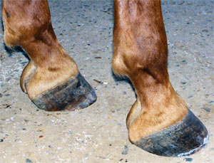
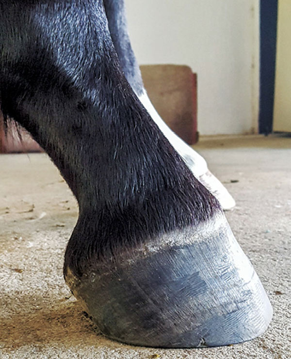
Club Foot Correction This horse is clearly standing under, ie unloading his painful heels Deformed boxy front hoof, long heels, curved coronet and short toes 8 months later – square stance;Of club foot A horse with club foot has one hoof that grows more upright than the other The "up" foot is accompanied by a broken forward pastern, that is, the hoof is steeper than the pastern (Photo 1) In a normal foot, the hoof capsule and the pastern align Radiographs willRadiography, Xray, Foot Cost $100 to $500 These cost ranges are approximate and may vary from region to region Additional charges may also apply Summary Radiographs of the equine foot or feet are a standard diagnostic used for hoof or foot related problems and in lameness exams, because a high percentage of lameness originates in this
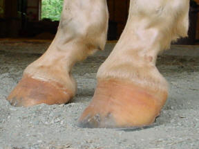
Club foot refers to a hoof that is more upright than normal It is often associated with a concave front (dorsal) hoof wall, high (often contracted) heels, and widening of the white line from mechanical stretching of the hoof wall attachments (the laminae) Adult club foot requires a completely different approach to treatment than juvenile club footMy horse was xrayed in the front and she was diagnosed as having club feet in the front, and perhaps the rear One of her ear feel turns out while standing square I was told that as she ages into her mid teens, her coffin bone would cause pain and she would then need to be put down She is 910 now I use her for team roping A study looking at 72 horses that underwent MRI for recent onset of navicular syndrome but without abnormalities detected on radiographs found the following 62 horses (86%) abnormalities in the navicular bone 32 horses (44%) pathologic changes of the DDFT 54 horses (75%) pathologic changes of the CSL
Diagnostic Imaging in horses At Sport Horses Vet we have portable equipment such as Xrays on high resolution horses, this helps us to reach a diagnosis to provide our customers with a high quality service, which allows us to obtain results in the act of great advantage for the horse vets and without the need to mobilize the animal This technology is mainly used for horses vet in theClub foot horse x ray Almost always, I am called to club foot cases when the horse goes lame on the normal side This is usually caused by people trying to force the hooves to match each other This thinking often leads a farrier to cut the sole out from under P3 at the toe and allow the heels to grow unchecked on the normal foot TwoAs of this 24year old horse has a Suspensory Ligament Issue on a club foot With the photo on the left the horses Suspensory Ligament is under strain, to aid in relieving some of this tension the heels were trimmed down This is a work in progress Egg bar Shoe Used for treating horses with diseases of the leg


Recognizing And Managing The Club Foot In Horses Horse Journals


Understanding Club Foot The Horse Owner S Resource
The Equine EZ Block is the best time saving tool a veterinarian has for getting Lateral & D/P (or A/P) Radiographs of both the Horse’s FRONT and HIND Feet Nothing is more frustrating than getting a horse's foot on an xray block, getting the cassette ready and having them step off Club foot is one of the most common deformities in the horse world Horses affected with club foot develop a flexural deformity of the coffin joint, due to a shortening of the musculotendinous unit that starts high up in the limb and inserts on the coffin bone in the foot, resulting in an upright conformation of the foot A club foot alters a horse's hoof biomechanics, frequently leading to secondary lamenesses Affected horses tend to land toefirst, and their heel's growth rate is


2


Boarding At Yucca Veterinary Medical Center
Plantar flexion at the ankle In the horse, hoof growth is dictated in large part by weight distribution If a horse puts more weight on the inside of a hoof, the blood is pushed to the opposite side of the foot causing faster growth and wearing down the weighted surface at a faster rate With respect to the club foot, the heel of the affected foot grows faster and the hoof The condition of the foot, the way the horse stands and your shoe modification ability will help determine the end result With all this in mind we worked on a club foot case recently This particular horse, a six year old gelding, has what I feel is a grade three club foot (on a 15 scale) Apparently the club foot condition has been


Michael Porter Equine Veterinarian November 12


Hoof Evaluation Radiographs For The Farrier
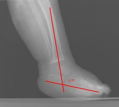
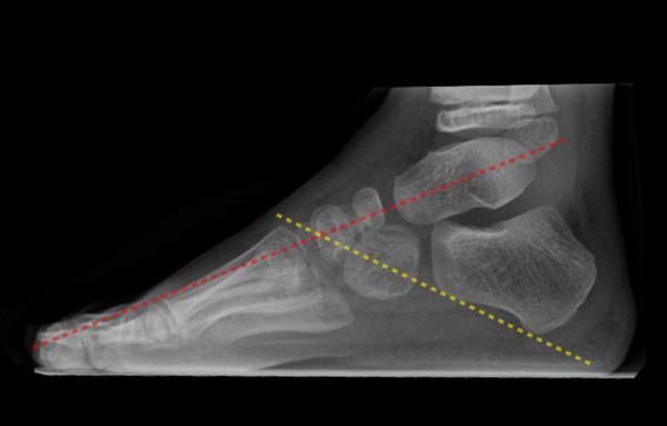
Club feet Very common abnormality possibly related to in utero positioning Calcaneus in equinus (pointed down) Parallel axes of talus and calcaneus on botThe name of the view describes the direction of the xray beam The beam is aimed from dorsoproximal to palmarodistal at a 65 degree angle to the sole of the foot This view may be obtained with the horse standing on the cassette as in this illustration The xray beam is centered at the coronary band Notice in the photo that the cassetteMost reliable roentgenographic view is the lateral projection, usually with the foot in maximum dorsiflexion in clubfoot there is no convergence of talocalcaneal region (parallel alignment), and the tibiocalcaneal relationship reveals equinus;


Hoof Evaluation Radiographs For The Farrier


Understanding X Rays The Laminitis Site
Post by sophielouise on at 304am When Chester had both front feet xrayed at 'home' (numerous 'takes', if you like) I think the xrays were about £150 It must have been a mobile unit I think, as they came out to the yard and set up in a spare box and the Xrays were instant on a laptop Horses with navicular disease typically have "classic" radiograph findings, making diagnosis fairly straightforward Veterinarians recommend taking a minimum ofIn infancy, ossification centers of the talus, calcaneus, and cuboid are present, whereas the navicular does not appear until 34 yr;


2


Contact Us Palmetto Equine Veterinary Services
A horse with an upright alignment of the pastern bones will also have upright hoovesa situation that is sometimes mistaken for club foot A true club foot is significantly more upright than the other hooves, or the angles of both hoof walls are steeper than the angles of the pasterns The severity of the problem is commonly graded on a four Understanding hoof Xrays Horse & Hound 31 July, 06 1237 Diagnostic techniques For many years, Xrays have been the major imaging technique for evaluation of the foot, for both diagnosis andAn X ray of your horse's foot can help you predict the future while it shows you the present MECHANICAL LAMINITIS TREATMENT Foot X Rays A Crystal Ball?


Fighting Laminitis Expert How To For English Riders


Equine Podiatry Say What Mobile Veterinary Services
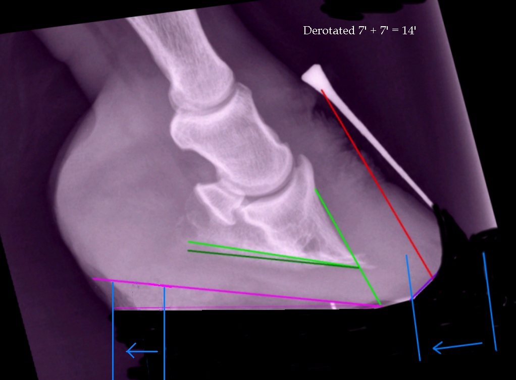
Positioning the foot for the examination Blocks are needed to elevate the off the ground allowing the foot to be centered in the cassette and the xray beam to pass horizontally through the specific area of interest (ie solar surface of the foot, DIP joint, navicular, etc) The foot should be placed as close the inside of the block whenThe search terms (horse* OR equine*) AND (foot OR feet OR digit* OR hoof OR hooves OR phalan * OR navicular) AND (radiograph* OR radiolog *) were generated and input into the PubMed search engine Following exclusion of studies more than 5 years old and those determined not to relate directly to the question, six useful results were yieldedComes very useful in horses with upright feet, the best example being the clubfooted horse As the foot grows out in these horses, there is a propensity for the dorsal wall to distort and flare, producing multiple angles to the dorsal wall Radiographic evaluation of the dorsal wall with a conforming marker allows accurate assessment of the


Equine Podiatry Dr Stephen O Grady Veterinarians Farriers Books Articles


Hoof Evaluation Radiographs For The Farrier
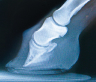
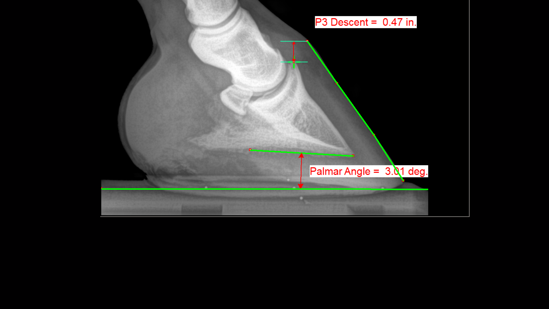
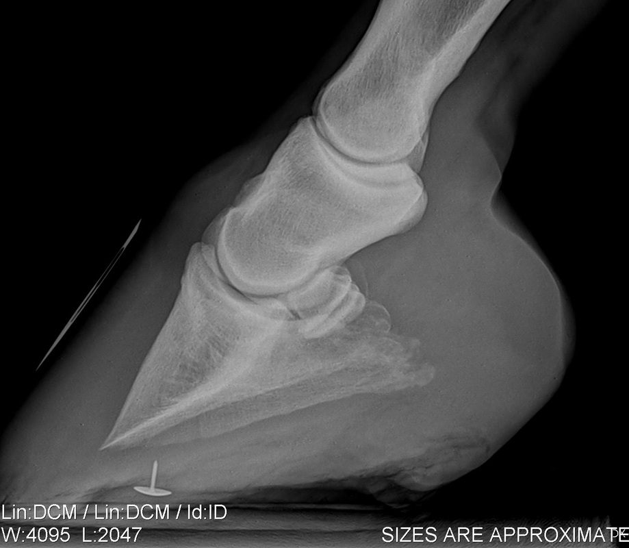
Same foot – 8 months later Pedal Bone Protrusion in a 5 yearold before trim with shoes on;In the club foot because the deep flexor tendon is contracted, the xray will show that the pedal bone angles are quite different, the front is not in line with the hoof wall, the tip is pointing down and the rear part is much greater than five degrees Put simply, the heels will need to be lowered and any flare corrected at the toe Introduction of Club Foot Club foot is also called talipes equinovarus It is a congenital deformity of the leg occurring in 1 or 2 of thousand births Males are more affected Male female ratio is 21 One third of these cases are bilateral CAUSES of club foot Actual cause of club foot is still unknown But the hypotheses are Genetic


Introduction To Clubfoot Physiopedia


High Heels The Laminitis Site
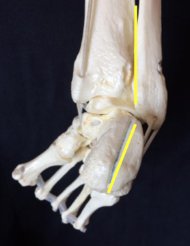
• rotatory subluxation of talocalnoenavicular joint (subtalar) complex with talus in plantar flexion and subtalar complex in medial rotation and inversion • also reffered as clubfoot • talipes derived from term talus ankle & pes foot • equinovarus derived from word equino like a horse & varus turned inward 4It might be a horse with very distorted feet, or a specific pathology that muddies the waters a bit In these cases, hoof radiographs (xrays) can be quite enlightening The information a well taken hoof radiograph can give you is tremendous, especially with pathology or severely distorted feetXray Scroll Stack Scroll Stack Lateral Lateral radiograph of the right foot shows that the long axes of the talus and calcaneus are nearly parallel The longitudinal arch is abnormally high AP radiograph of the right foot shows abnormally narrow talocalcaneal angle, with severe adduction and supination of the forefoot


Filly With Club Foot Barefoot Hoofcare


Filly With Club Foot Barefoot Hoofcare
Too much bad stuff and you have an unusable horse You take side xrays to determine the degree of rotation, and of separation (if any) of the coffin from the hoof wall You take bottom xrays to determine the degree of chipping/degeneration in the leading edge of the coffin bone If your vet is on the fence about whether the horse has an abscess or not they may decide to xray the foot, which can show an abscess or possible other issues causing the horse to be lame This xray of the foot shows a dark circular lesion on the right side, which is consistent with an abscess w/ club foot, axes of talus & calcaneus becomes more parallel;


Equine Podiatry In Wendell Nc Neuse River Equine Hospital


The Wild Mustang Hoof
In the scenario where the DP is ordered to query a foreign body, do not angle the xray beam to mimic the arch as this will result in elongation of the foreign body in question In trauma, the patient may not be able to flex the affected knee to the desired angle In this case, a triangular wedge can be placed under the footMethod of Beatson and Pearson AP of Foot taken in 30 deg plantarflexion, with the xray tube directed 30 Figure 1 is the affected foot and figure 2 is the normal foot Xray vision was not necessary in this case to confirm the diagnosis due to the classic distortion of the hoof capsule Club feet in foals develop from tendon contracture or secondary to accelerated skeleton growth


Hoof Care For The Club Footed Horse David Farmilo


Basic Shoeing Working With A Club Foot Farrier Product Distribution Blog
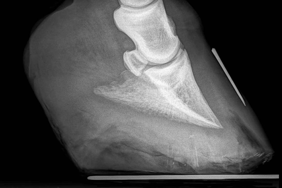
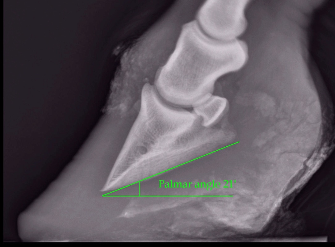
I talked to the vet, he said the xrays of the feet were good with a bit of "rust" in the joints but the horse was sounder than most 5 year olds and would last a long time with good care The farrier I'm using initially trimmed the horse (was told he could go without shoes), but he was ouchyThe xray will show whether the hoof pastern axis is parallel If the axis is broken forward (club foot) or if the axis is broken back (long toe underrun heel), the radiograph will reveal the degree of deformity and the best way to trim the foot to improve it Using landmarks, measurements can be drawn on the radiographs and transferred to the Talocalcaneal parallelism is the radiographic feature of clubfoot (talipes) Simulated weightbearing xrays are used for infants who have not commenced walking Positioning for foot xrays is very important The anteroposterior (AP) view is taken with the foot in 30° of plantarflexion and the tube at 30° from vertical


Recognizing Various Grades Of The Club Foot Syndrome


1
Top view xray of foot Note the talus and navicular bones (two sets of green arrows) badly offset as if the navicular were pulling off the talus The red arrows point out three different stress fractures in the metatarsals caused by mere walking The xrays showed fractures in the lateral cartilage (or sidebones) on both front feet The vet said these fractures were due to the extreme pressure put on the cartilage (due to the size and placementof the sidebone) and also the lack of shock absorption (due to heel contraction and size of the hoof)Radiographic Assessment of Club Foot Discussion helps to determine preoperative pathology;


Equine Laminitis Soft Ride Boots For Equine Laminitis Comfort


Pro Equine Grooms Contracted Heels In Horses
Congential Talipes equinovarus it`s a common form of clubfoot Talipes = talus (ankle) pes (foot) Equino = heel is elevated (like a horse's) varus = turned inward With this type of clubfoot, the foot is turned in sharply and the person seems to be walkingon their ankle 3 Clubfoot, or talipes equinovarus, is a congenital deformity consisting of hindfoot equinus, hindfoot varus, and forefoot varusThe deformity was described as early as the time of Hippocrates The term talipes is derived from a contraction of the Latin words for ankle, talus, and foot, pesThe term refers to the gait of severely affected patients, who walked on their anklesXray of feet (typical clubfoot) Clubfoot Introduction Clubfoot (talipes equinovarus – TEV) is one of the major orthopedic conditions of childhood One of the most common of all birth defects, clubfoot affects about 1 in 400 babies born in the United States each year Boys are affected twice as often as girls Causes


Low Foot Case Study Dixie S Farrier Service


Recognizing Various Grades Of The Club Foot Syndrome
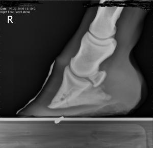
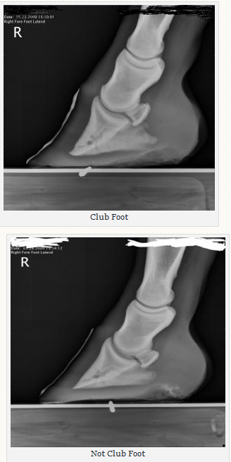
Club Foot Not Club Foot These are XRays of the front feet of a yearling filly The first figure is the right foot, the bottom is the left The top photo depicts a classic clubfoot, the bottom is a normal foot The external evidence indicating it is a clubfoot is the curved, dished wall of the foot The coffin joint angle is the radiographicXray is one of the most widely used forms of diagnostic imaging in both human and veterinary medicine At the RVC it is a regular part of our work to use x


Comstock Equine Hospital Veterinarian In City State Country Corrective Trimming


Michael Porter Equine Veterinarian November 12


Low Foot Case Study Dixie S Farrier Service


Club Foot Or Not Barefoot Hoofcare


Puncture Wounds In The Foot Vetsouth


Podiatry Burwash Equine Services


Viewing A Thread Ringbone Help


Correct


Recognizing And Managing The Club Foot In Horses Horse Journals


So Called Club Foot By James R Rooney Dmv


Hoof Deformities Round Pen Square Horse


The Truth About Hoof Pastern Axis


Thrush Cured Cowboy Pads And Copper Sulfate


Clubfoot Imaging Practice Essentials Radiography Computed Tomography


Understanding X Rays The Laminitis Site


How To Treat Club Feet And Closely Related Deep Flexor Contraction


Severe Lameness Scarsdale Vets


Laminitis Signalment Treatment And Prevention Flying Changes


2


2


The Progressive Equine Services Hoof Care Centre Facebook


2


Shoeing Options For Club Foot In Horses


Equine Therapeutic Farriery Dr Stephen O Grady Veterinarians Farriers Books Articles


Radiographic Exposure Settings Hints And Tips Equine Imv Imaging


Recognizing Various Grades Of The Club Foot Syndrome


Frequent Trips Aid Club Foot American Farriers Journal


2


Does My Horse Have Pyramidal Hoof Disease The Horse


Foal With Hoof Problems Club Foot The Horse


Club And Subluxation In The Proximal Interfalangian Articulation Farriers Forum


Clubfoot Barefoot Hoofcare


Recognizing Various Grades Of The Club Foot Syndrome


Club Foot Or Upright Foot It S All About The Angles American Farriers Journal


Epublishing


Recognizing And Managing The Club Foot In Horses Horse Journals


Shoeing Options For Club Foot In Horses


The Importance Of Physical Maturity In The Horse Horsetalk Co Nz


2


2


Podiatry The Equine Center


Understanding X Rays The Laminitis Site


Natural Angle Volume 15 Issue 1 Spanish Lake Blacksmith


Farriery For The Hoof With A High Heel Or Club Foot Semantic Scholar


Understanding X Rays The Laminitis Site


Radiography Of The Equine Stifle Part 4 Of 4 Imv Imaging


Talipes Equinovarus Clubfoot Radiology Case Radiopaedia Org


Club Foot


Equine Therapeutic Farriery Dr Stephen O Grady Veterinarians Farriers Books Articles


Managing The Club Hoof Easycare Hoof Boot News


Club Foot In Horses Brian S Burks Fox Run Equine Center Facebook


The Wild Mustang Hoof


Congenital Talipes Equinovarus Or Club Foot Bone And Spine


Recognizing And Managing The Club Foot In Horses Horse Journals


Club Foot Or Upright Foot It S All About The Angles American Farriers Journal


1


2


Understanding X Rays The Laminitis Site


Frequent Trips Aid Club Foot American Farriers Journal


1


2


Types Of Crooked Legs In Foals


2


The Chronicle Of The Horse


Understanding Club Foot The Horse Owner S Resource


2


Radiolucency Glue On Horseshoes By Sound Horse Technologies


Radiographic And Radiological Assessment Of Laminitis Sherlock 13 Equine Veterinary Education Wiley Online Library


Pdf Evaluation Of Club Foot In Working Donkeys


Recognizing And Managing The Club Foot In Horses Horse Journals


Hoof Radiographs Springhill Equine Veterinary Clinic


Hot Topics In Hoof Care Part 4 The Abnormal Horse Hoof The Horse


Recognizing And Managing The Club Foot In Horses Horse Journals


Innovative Equine Podiatry Navicular Case Examples


Equine Therapeutic Farriery Dr Stephen O Grady Veterinarians Farriers Books Articles


How To Manage The Club Foot Birth To Maturity The Horse
コメント
コメントを投稿